Kirichenko Margarita |
 |
Faculty CITA | |
Group: CSD 01-b | |
Scientific adviser: Candidate of Technical Sciences Omelchenko A.A. | |
Master's Degree work: | |
"Models and algorithms of the dedicaded computer |
| Autobiography | Links | Individual task | Library | Report about the search |
Thesis to master's degree work
In russian language
to do analysis of methods for focusing optical instruments which are used nowadays;
to define criterias of high-quality image;
to select test object on the picture of blood smear;
to create the model for calculation of characteristic for test object;
to develop the algorithm for focusing microscope (define direction of lens movement);
to create software for this algorithm.
Creation of the «Fokus-mikro 1.0» system will allow to get the high-quality image of blood smear, to minimize investigation time and also to improve working process for operator. It will help to reduce eye tension because of no need to look at the lens for a long time.
page up
Microscope focusing is the task about abject detection.It is necessary to distinguish good-focusing image from the image with well
sfokusirovannoe image of object from the same object with degraded ages. The object on the blood smear is conditional point on the image - bacterium. It is different from surrounding because of deeper color. There is a big difference between the object which we are interested in and and its environment. On this So on the
first step it is necessary to recognise the bacterium (listed below as conditional point). For analysis it is offered a set of images from the one blood smear which has been gotten at the different focal distance (i.e. at different position of microscope makro- and mikroscrews). For
all conditional points which were found on every image, it is offered to do analysis based
on three features:
- total area of the object;
- middle (or integral) brightness;
- contrasting on the object contour.
It is necessary to determine the edge of conditional point (cell) for estimation of these characteristics.
Making SСS for focusing microscope it is necessary to pay attention for some factors. Above all things note should be taken on
that in swingeing majority of image, got by means of microscope are characterized by the presence of noise arising up from uneven illumination
insufficient cleaning of laboratory equipment, own noises fotopriemnih devices and other reasons. At the automated analysis it is necessary to take into
account and to compensate the presence of noise. Otherwise influencing of noise can distort
result.
page up
Review of existent researches and developments
The digital image processing is the independent region of knowledge, which quickly develops in many countries. With the image
processing it is necessary to have business to the specialists of a different type. On questions from this region a lot
of works and literatures are published, for example, Oppengeym A.V., Shafer R.V. «Digital treatment of signals», Soyfer V.A.
«Computer image processing» and dr. The MatLab package is equipped in a number of functions allowing to produce treatment
of signals and images.
For the image processing вейвлет-transformations which became a necessary mathematical instrument in many researches are
used also. In particular, they can be used for focusing of microscope. It is comparative easy task for veyvletov, because
the calculated вейвлет-coefficients are great at the well sfokusirovannom image and strongly fall at defokusirovke microscope.
At some level of permission, proper to the natural scale of image of object, this effect of defokusirovki becomes especially
strong. At other level he is expressed less brightly, but becomes more asymmetric depending on that, there is a microscope
higher or below than point of focusing. This property of asymmetry was used with the purpose of automation of process of
focusing of microscope, as it is clear sets direction of his motion toward focusing position. Such method can be successfully
applied in many other regions, rather than just in medicine.
page up
Purpose and tasks of master's degree work
Theme of master's degree work – «Models and the SСS
algorithms focusing of microscope». The SСS planning will be conducted on a base
research laboratory of NII of medical problems of family.
The purpose of the given project
is:
- obektivizatsiya process of receipt high-quality
images for the decision of the put task;
- abbreviation of time of receipt of high-quality picture
bloods;
- improvement of terms of labour of operator, namely, decline
loadings on his visual vehicle due to the exception of necessity of protracted considerations of preparation through the eyepiece of microscope due to development
of models and algorithms of the automatic focusing of microscope, and creation on their
basis SСS.
For achievement of the put aims in master's degree work
it is necessary to decide the row of tasks: analysis of existent researches
and developments; determination of criteria of sfokusirovannogo image, on the basis of what is conducted
calculation of necessary descriptions and properties.
page up
Theoretical analysis and mathematical model of object of research
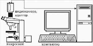
Picture 1. Chart of forming entrance data
The prepared preparation of blood is placed on subject table
microscope. At different position of makro- and mikrovintov of microscope get
row of pictures of the same preparation. For developed SСS is used video camera OSCAR, connected to the personal COMPUTER by videokarti of
the ASUS firm having the televisional entrance. For the capture of videoizobrageniya the program
is used ASUSLIVE, which forms the shot of image of stroke of blood in the format of
*.bmp and destroys him on the screen of monitor. Examples of the got images:


Picture 2.
Got images of strokes of blood
page up
Generalization of results of scientific search and analysis
As a result of implementation magistrskoy works were existent methods, algorithms and systems of focusing of microscope are considered by means of recognition of image. Their analysis allowed to define basic mathematical methods which are used for creation of specialized computer system for focusing of microscope on the pictures of strokes of blood, by a purpose which abbreviation of time and obektivizatsiya process of receipt is high-quality image. Effective ways and methods for the decision are found put task.
page up
Прэтт У.К. "Цифровая обработка изображений". - М.: Мир, 1982. T.I.-2. 792 с.
Рудаков П.И., Сафонов В.И. "Обработка сигналов и изображений". - М.: ДИАЛОГ-МИФИ, 2000. - 416 с.
Сойфер В.А. Компьютерная обработка изображений. Часть 2 Методы и алгоритмы // Соровский образовательный журнал, №3,1996
Хуанг Т.С. Обработка изображения и цифровая фильтрация. М.: Мир, 1979. 274 с.
Хорн Б.К.П. Зрение роботов. Москва "мир", 1989 г, 484 стр.
http://www.ksu.ru/nilkto/cell/rasdel1/r1_p3_s1.html
page up